Echocardiography Test
GET UPTO 50% OFF
Get upto 50% Discount At Your Nearby TEST CENTRE
60+ Test centres in India
Satisfaction Rate of More than 95%
Trusted By 30,000+ People
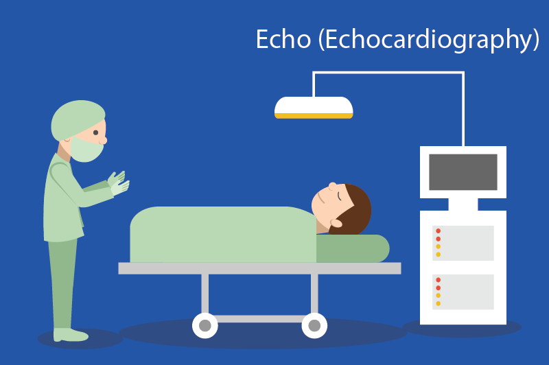
Echocardiography Test
Normally these structures need to be intact and the heart muscle needs to beat in a coordinated for the heart to function, so that blood flows in and out from each chamber in the proper direction.
The word echocardiograph means:
- Echo=sound
- Card=heart
- Gram=drawing
An electrocardiogram (EKG, ECG) is the most common heart tracing technique. It create moving pictures of your heart by using sound waves. The picture shows the structure and anatomy of the heart.

- The echocardiogram is like the other tests that are done to evaluate heart anatomy and function of the heart.
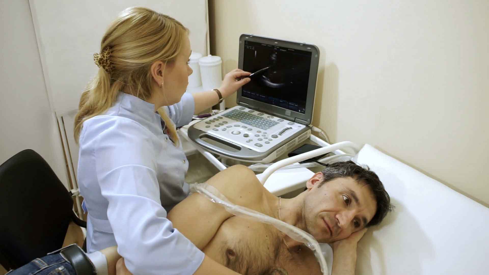
- An echocardiogram is an ultrasound test that is used to detect the structures of the heart, as well as the direction of blood flow within the heart.
- The images and videos are produced by the specially trained technicians.
- The electro cardiograph is done by using instruments like probe or transducer that is then placed in various places on the chest, to see the various anatomies of heart.
- Specially trained cardiologists views the report to provide the proper result of the test.
- The human heart is a two-stage electrical pump that helps to circulates blood throughout the body. Heart has four chambers and four valves.
The electrocardiograph test is used to recognize the function of your heart and also helps to detect the presence of many types of heart disease, such as pericardial disease, infective endocarditis, pericardial disease, infective endocarditis, valve disease, myocardial disease, , cardiac masses and congenital heart disease.
What happens during an Echocardiogram Test?

- Before the test, the procedure is explained by the specially trained cardiac sonographer, including their side effects.
- If you have any query then you also can discuss your questions regarding test with the cardiac sonographer.
- A cardiac sonographer will provide a gown to wear.
- A cardiac sonographer will ask you to remove your clothes from the waist up.
- Electrocardiograph (EKG) monitor contains 3 electrodes (small, flat, sticky patches) on your chest. The electrodes are attached to an electrocardiograph (EKG) monitor that that helps to prepare a charts regarding your heart’s electrical activity during the whole test.
- Sonographer will apply gel on the end of the electrodes, which will not harm your skin and the body.
- This gel helps produce clearer pictures.

Contact us for the best services
When you fill out this form, our executive will call you and book your time slot, so that your time is saved.